
Skin Cancer
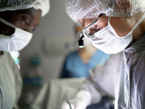
Skin Cancer: Diagnosis, Surgical Treatment, and Reconstruction
Skin cancer is one of the most common cancers and frequently occurs in areas exposed to ultraviolet (UV) radiation, including the face, scalp, ears, eyelids, nose, lips, neck, and hands. Plastic surgeons play an important role in the management of skin cancer by performing oncologic resections while preserving critical structures and restoring appearance and function through reconstructive techniques.
Types of skin cancer and surgical margins
Basal cell carcinoma (BCC)
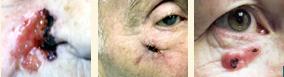 |
Basal cell carcinoma is the most common type of skin cancer. It usually grows slowly and has a low risk of spreading; however, untreated BCC can invade surrounding structures, particularly in high-risk areas such as the nose, eyelids, ears, and lips.
Treatment is primarily surgical excision with removal of the tumour and an appropriate margin of normal-appearing skin to reduce the risk of recurrence
Typical excision margins:
- Low-risk BCC: approximately 3–4 mm margins of clinically normal skin may achieve high rates of complete removal.
- High-risk BCC: wider margins or margin-controlled techniques such as Mohs micrographic surgery may be recommended, particularly for lesions on the central face, eyelids, nose, ears, lips, recurrent tumours, aggressive subtypes, or larger lesions.
Squamous cell skin cancer
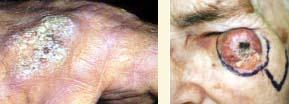 |
Squamous cell carcinoma has a greater potential than BCC to invade deeper tissues and spread, particularly when located on high-risk sites or in patients with immune suppression.
Typical excision margins:
- Low-risk SCC: approximately 4–6 mm margins are commonly recommended.
- High-risk SCC: wider margins or Mohs surgery may be considered depending on tumour characteristics, including size, depth, location, growth pattern, and pathology features.
High-risk features include:
- Location on the ear, lip, or central face
- Tumour size greater than 2 cm
- Increased depth of invasion
- Poorly differentiated tumour type
- Perineural invasion (spread along nerves)
- Recurrence after previous treatment
Malignant melanoma
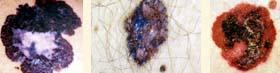 |
Melanoma arises from melanocytes, the pigment-producing cells of the skin. Although less common than BCC or SCC, melanoma can spread to lymph nodes and distant organs, making early diagnosis and treatment essential.
Diagnosis begins with recognition of suspicious lesions using the ABCDE melanoma warning signs:
- A – Asymmetry: One half of the lesion does not match the other half.
- B – Border: Irregular, scalloped, or poorly defined borders.
- C – Colour: Variation in colour, including shades of brown, black, red, blue, or white.
- D – Diameter: Lesions larger than approximately 6 mm (about the size of a pencil eraser) are more concerning, although smaller melanomas can occur.
- E – Evolving: Any change in size, shape, colour, elevation, symptoms, bleeding, or appearance over time.
A biopsy is required to confirm melanoma and determine tumour thickness, which is the most important factor guiding treatment.
Melanoma excision margins
Surgical treatment involves wide local excision with margins determined by the Breslow thickness of the melanoma:
- Melanoma in situ: approximately 5 mm margin of normal skin.
- Invasive melanoma ≤1 mm thickness: 1 cm margin.
- Melanoma 1–2 mm thickness: 1–2 cm margin.
- Melanoma >2 mm thickness: 2 cm margin.
For selected invasive melanomas, a sentinel lymph node biopsy may be recommended to assess whether cancer cells have spread to regional lymph nodes.
Reconstruction after skin cancer removal
Following complete tumour removal, reconstruction aims to restore both function and appearance. The reconstructive approach depends on the size, depth, and location of the defect.
Common techniques include:
- Primary closure: Direct repair of smaller wounds when surrounding tissue allows safe closure.
- Skin grafts: Transfer of skin from another area to cover defects when local tissue movement is not appropriate.
- Local flaps: Movement of nearby tissue with its blood supply into the defect, commonly used for facial areas such as the nose, cheek, eyelid, and lip.
- Complex reconstruction: Larger defects may require cartilage support, staged procedures, or microsurgical techniques to restore three-dimensional structure and function.
Special Considerations in Facial Skin Cancer
Facial skin cancers require careful planning because small areas often contain critical structures.
- Eyelid reconstruction: Must restore eyelid closure, protect the eye, and maintain vision.
- Nasal reconstruction: Must preserve nasal contour, breathing function, and facial balance.
- Lip reconstruction: Must maintain oral competence, speech, sensation, and appearance.
- Ear reconstruction: Must preserve the complex shape and framework of the ear.
Prevention and Follow-Up
Early detection improves outcomes. Patients should perform regular skin examinations and seek medical assessment for any changing or concerning lesion. Sun protection, including regular sunscreen use, protective clothing, and avoidance of excessive UV exposure, remains an important part of prevention.
Plastic surgeons work closely with dermatologists, pathologists, and oncology specialists to provide comprehensive skin cancer care—from diagnosis and oncologic excision to reconstruction and long-term surveillance.